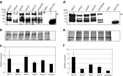
Fig. 9.
Effect of deletion of ERAD components on the amount of GlaGus fusion protein in total protein extracts. Western analysis of GlaGus amounts in total protein of mycelium samples of scGlaGus (a) and mcGlaGus (d) ERAD deletion strains. Samples were grown in CM for 24 h at 30°C. Ten micrograms of total protein was separated by gel electrophoresis and immunodetected with an anti-Gus antibody. Detection was carried out through a chemiluminescence reaction for 5 min. As a positive and negative control, 50 ng of purified Gus and a total protein extract from N402 were loaded. The arrow indicates the band corresponding to the GlaGus fusion protein (≈140 kDa). The relative amounts of protein were normalized for loading differences by comparison with a “twin” gel stained with Coomassie blue (b, e). c, f Relative amount of GlaGus fusion protein detected in total protein extracts of strains with impaired ERAD and respective parental strain. Bars indicate standard deviations from two independent experiments

