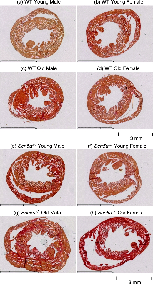
Fig. 5.

Representative slide for cardiac fibrosis staining in WT and Scn5a +/− hearts grouped by age and sex. The study population was stratified into: WT young male (a), WT young female (b), WT old male (c), WT old female (d), Scn5a +/− young male (e), Scn5a +/− young female (f), Scn5a +/− old male (g), and Scn5a +/− old female (h). Hearts were routinely stained with Sirius red, and morphometric analysis for percentage of fibrosis was performed for all eight groups. Areas of increased red uptake signify presence of fibrotic changes. Horizontal bar below sections in each panel denotes a 3mm distance
