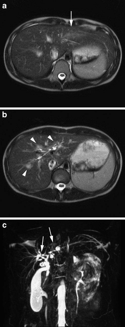
Fig. 15.
MRI and MRCP in a 10-year-old girl with ulcerative colitis and signs of cholestasis. a, b Axial rt TSE T2-W images show a wedge-shaped area of T2 hyperintensity (arrow in a) as well as periportal parenchymal hyperintensities and biliary duct dilatations (arrowheads in b). c MIP reconstruction of the rt 3-D TSE T2-W sequence clearly illustrates the dilatations and strictures in the common bile duct and central intrahepatic bile ducts (arrows) compatible with primary sclerosing cholangitis

