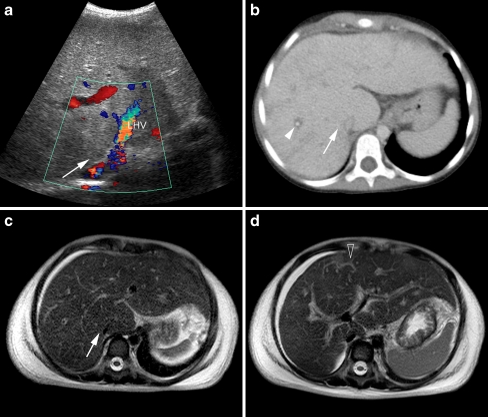
Fig. 19.
US, CT and MRI in a 8.5-year-old girl presenting with acute abdominal pain and distension. a Colour Doppler US shows heterogeneous hyperechoic liver parenchyma with absent flow in the main hepatic veins of the right hepatic lobe (arrow), LHV left hepatic vein. b Axial CT slice shows slightly heterogeneous enhancement and periportal oedema (arrowhead) as well as absent enhancement of the right hepatic veins (arrow). c and (d) rt TSE T2-W axial MR images show heterogeneous liver parenchyma, ascites, absent right hepatic veins (arrow in c) and intrahepatic collaterals (open arrow in d), compatible with Budd-Chiari syndrome

