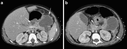
Fig. 20.
Contrast-enhanced CT in a 3-year-old boy with acute lymphatic leukaemia and acute pancreatitis most likely related to Asparaginase (chemotherapy). The CT shows diffuse gland enlargement with mild inhomogeneous enhancement of the parenchyma (open arrow in a), irregular margins and inflammatory changes of the peripancreatic tissue (arrowhead in b), and multiple small and large peripancreatic fluid collections (arrows in a). The inflammation has spread anteriorly to the pararenal space with thickening of Gerotha’s fascia and peritoneum (arrow in b)

