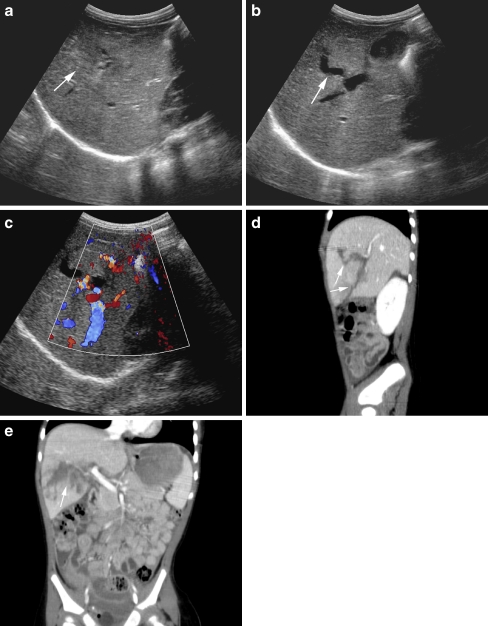
Fig. 22.
US and CT of the liver in a 1.5-year-old girl with a liver laceration due to blunt abdominal trauma. a–c US shows a heterogeneous defect in the liver parenchyma (arrow in a) with anechoic parts (arrow in b) extending into the liver hilum (c). The sagittal (d) and coronal (e) MPR reconstructions of the contrast-enhanced CT better illustrate the full extent of the liver laceration (arrow in d and e) caudal in the right liver lobe reaching into the liver hilum but without evidence of major vascular injury (grade 4)

