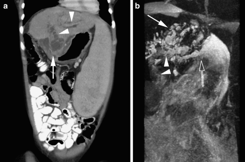
Fig. 5.
Images in a 19-year-old woman with a history of biliary atresia and Kasai procedure (hepatic portoenterostomy), now presenting with sepsis and cholangitis. a Coronal MPR of the CT scan of the abdomen shows massive splenomegaly, irregular dilatation of the biliary tree (arrowheads) and a complex multiloculated fluid collection (biloma) with the suggestion of a connection with the biliary tree (arrow). b MIP reconstruction of the rt 3-D TSE T2-W sequence also shows the portoenterostomy (arrowheads), irregular dilatation of the biliary tree (closed arrow) and the close relation of the biliary tree to the biloma (open arrow). Furthermore, there is ascites visible surrounding the liver

