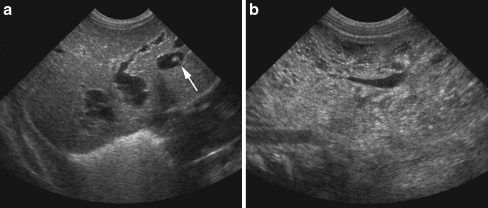
Fig. 8.
Antenatally diagnosed liver cysts and enlarged kidneys in a 4-day-old boy. a US of the liver shows multiple fusiform and cystic dilatations of the intrahepatic bile ducts. One of the cysts shows the “central dot” sign, representing the portal fibrovascular bundle (arrow); type V choledochal cysts. b US of the kidneys shows enlargement and increased echogenicity with multiple small cysts characteristic of polycystic kidney disease; Caroli disease

