
Figure 2.
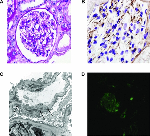
Renal transplant biopsy. (A) Light microscopy showing a normal glomerulus (periodic acid-Schiff). (B) Immunoperoxidase staining showing granular capillary wall and mesangial C3 deposition. C3 was detected using a polyclonal rabbit anti-human C3c antibody (Dako Ltd., Ely, United Kingdom). (C) Electron micrograph showing subendothelial deposits (denoted by arrows) with new basement membrane beneath. (D) Glomerular C5b-9 by indirect immunofluorescence. C5b-9 was detected using a monoclonal mouse anti-human C5b-9 antibody (DakoCytomation Ltd., Ely, United Kingdom) and a fluorescein isothiocyanate-labeled goat anti-mouse IgG (Sigma-Aldrich Company Ltd., Dorset, United Kingdom).