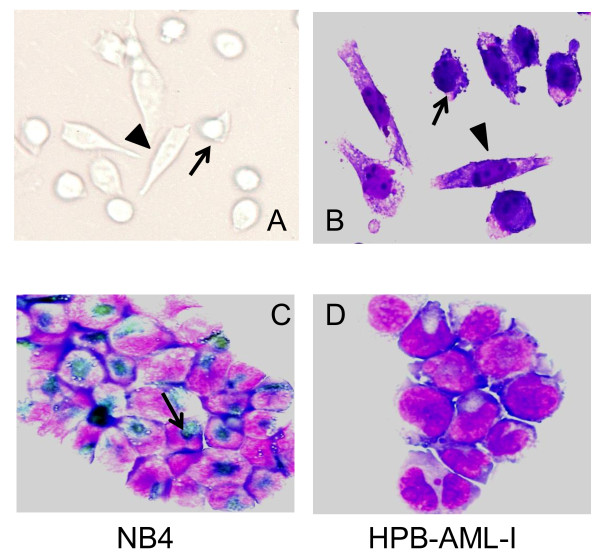
Figure 1.
Morphological and cytochemical characteristics of HPB-AML-I. Inverted microscopic examination (A) and May Grünwald-Giemsa staining (B) revealed that HPB-AML-I features a round-polygonal (arrow) and spindle-like (arrowhead) morphology. The human acute promyelocytic leukemia (APL) NB4 cell line was used as positive control for myeloperoxidase staining. Positive reactions are indicated with an arrow (C). Absence of myeloperoxidase expression was observed in the cytospin-prepared HPB-AML-I cells (D). Original magnification ×400.

