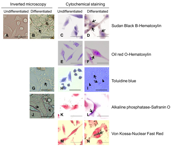
Figure 4.
Morphological and cytochemical changes in HPB-AML-I cells following the induction of differentiation toward mesenchymal lineage cells. Undifferentiated HPB-AML-I cells observed with an inverted microscope are shown for comparison (A). A representative HPB-AML-I cell induced to differentiate toward adipocyte and showing spindle-like morphology and cytoplasmic vacuoles is indicated with an arrow (B). Undifferentiated (C, E) and differentiated (D, F) HPB-AML-I cells were stained with Sudan Black B (C, D) and oil red O (E, F). The nucleus was counterstained with hematoxylin. Positive Sudan Black B and oil red O staining of cytoplasmic vacuoles of the differentiated HPB-AML-I cells is indicated with an arrow. Following the induction of differentiation toward chondrocytes, HPB-AML-I cells showed polygonal morphology with a number of cytoplasmic vacuoles (arrow) (G). The micromass of undifferentiated (H) and differentiated (I) HPB-AML-I cells were stained with toluidine blue. The presence of lacunae (arrows) and the toluidine blue-positive extracellular matrix (arrowheads) characteristic for a cartilage were observed following the induction of chondrogenesis. The osteogenic-differentiated HPB-AML-I cells demonstrated a number of cell processes (arrow) and an eccentrically located nucleus (arrowhead) (J). Undifferentiated (K) and differentiated (L) HPB-AML-I cells were cytochemically examined for alkaline phosphatase expression. The nucleus was counterstained with Safranin O. Positive reactions are shown in the differentiated HPB-AML-I cells with an arrow. Undifferentiated (M) and differentiated (N) HPB-AML-I cells were stained with von Kossa method. The nucleus was counterstained with nuclear fast red. The extracellular depositions of calcium following the induction of osteogenesis are indicated with an arrow. Original magnification x400; Size bar: 20 μm.

