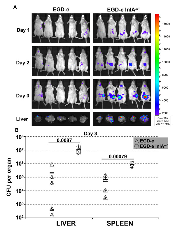
Figure 8.
Bioluminescent imaging (BLI) of Balb/c mice orally infected with either EGD-e or EGD-e InlAm* (tagged with pIMK2lux). A. Balb/c mice (five per group) were gavaged with a total of 5 × 10⁹ cfu and the progression of infection in each mouse (labelled 1 thru 5) followed on day one, two and three by BLI. Pseudocolor overlay represents the light emission profile from the infected mice with the scale bar on the right hand side. On day three mice were euthanized and livers examined ex vivo by BLI. B. Total bacterial loads from livers and spleens were numbered. The cross line denotes the mean organ cfu recovery for the five mice. Statistical analysis was conducted using a student t test with the p-value shown on the graph.

