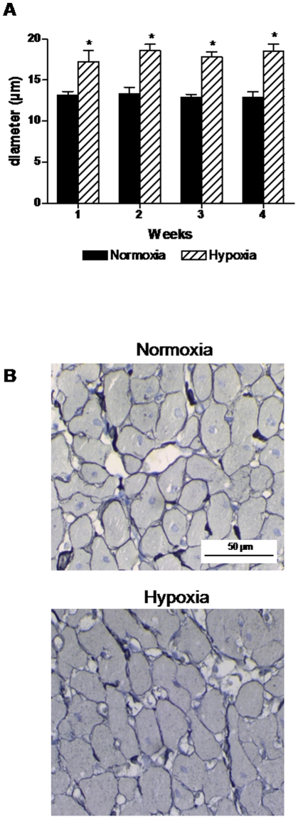
Figure 2. Morphometric measurements of the right ventricle.
A: cardiomyocyte diameter was significantly increased by hypoxia compared to normoxic controls. B: examples of staining with reticulin in right ventricle samples from normoxic and hypoxic rats. n = 5–6 in both groups at all time points. Values are means ± SE. *P<0.05 vs. normoxia at same time point.

