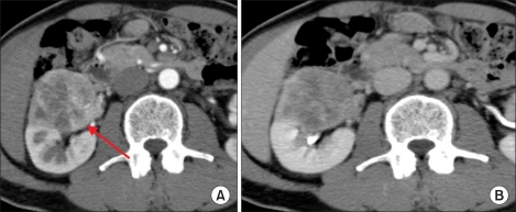
FIG. 2.
Preoperative contrast-enhanced CT images of the right renal tumor. The tumor was heterogeneously enhanced, and the degree of enhancement was weaker than the normal renal parenchyma in the early phase (A). The tumor showed mild wash-out in the delayed phase (B). We also found right renal venous invasion (A).

