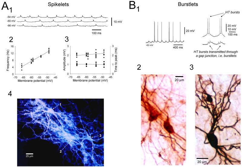
Figure 6. Evidence for GJ coupling between LGN TC neurons.
A. Rhythmic spikelets in a cat LGN TC neuron at different levels of injected d.c. current in the presence of 100 μM trans-ACPD. The frequency of these events is altered by membrane polarization (2) but the properties of individual events, i.e. amplitude (■) and time to peak (●), are not (3). This suggests that spikelets represent action potentials from distinct neurons which have been transmitted through a gap junction (GJ). This is confirmed by noting that injection of dye into this neuron leads to the staining of two additional cells (i.e. dye coupling) (4). Note that the ability to alter the frequency of firing in a TC neuron by varying injected d.c. current in a separate coupled neuron is suggestive of sparse coupling (see Hughes et al., 2004). Sparse coupling is also implied by the general lack of dye transfer to more than one or two additional cells (Hughes et al., 2002a). B1. LGN TC neuron exhibiting bursts of spikelets or burstlets (left trace). Note the intermittent entrainment of neuronal firing by these burstlets at this particular level of depolarization (cf. Fig. 4C; see also Fig. 8A in Hughes et al., 2004). The traces on the right show the similarity in properties of two consecutive HT bursts obtained from a different neuron (top) and two consecutive burstlets enlarged from the recording on the left (bottom). B2 and B3. Examples of dye coupling obtained following injection of markers into TC neurons exhibiting burstlets (B3 corresponds to the recording shown in Fig. 10). (A modified from Hughes et al., 2002a with permission from Elsevier; B modified from Hughes et al., 2004 with permission from Elsevier).

