
Figure 2.
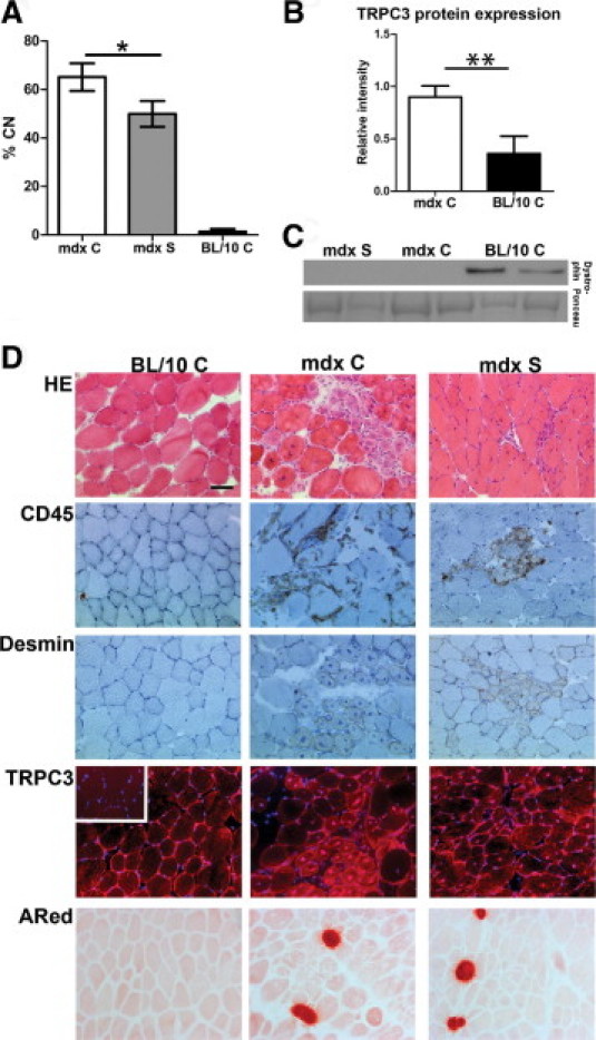
Treatment with streptomycin improves central nuclei counts and pathology in TA of mdx mice in utero and 6 weeks postnatal. A: Central nuclei counts (% CN) in C57/BL10 (BL/10 C), C57/BL10-mdx control (mdx C), and streptomycin-treated (mdx S) mice. Data are expressed as means ± SD of 3–5 mice. There is a statistically significant reduction in %CN in mdx S, compared with mdx C (*P < 0.05). B: Western blot detection of TRPC3 (mol. wt. ∼97 kDa) in crude muscle extracts. Ponceau stain band mol. wt. 80–90 kDa was used as standard and loading control. We detected significantly increased TRPC3 protein in mdx skeletal muscle, compared with BL/10 control (**P < 0.01). C: Western blot results of dystrophin (mol. wt. ∼427 kDa) expression in TA from 6-week-old mdx S, mdx C, and BL/10 C. There is no dystrophin protein expressed in mdx mice, regardless of treatment, indicating that read-through of the dystrophin stop codon mutation has not occurred. Representative results from two mice per group are shown. D: Representative images of histological examination of BL/10 C, mdx C, and mdx S mice. The H&E staining shows general histology; CD45 detects all lymphocytes and is a marker of inflammation; and desmin is expressed by regenerating and newly forming fibers and is a marker for immature fibers and ongoing regeneration. Overall histology points toward less necrosis and inflammation in mdx S, compared with mdx C. TRPC3 protein expression is detectable in membrane, cytoplasm, and nuclei of both mdx C and mdx S mice and the expression appears to be increased in newly formed myotubes. This TRPC3 expression pattern in mdx control and treated mice, compared with BL/10 C, correlates with the increased expression seen on Western blots (B). Inset: Negative control image for TRPC3 immunohistochemical staining for BL/10 C. Nuclei are counterstained with DAPI in blue. Alizarin Red (ARed) staining shows necrotic foci with calcium accumulations and there is no detectable difference between mdx C and mdx S. Scale bar = 50 μm.