
Figure 4.
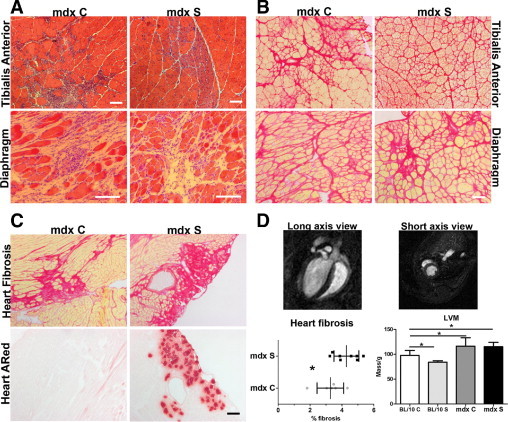
Improvement of limb muscle pathology combined with aggravation of cardiac pathology after continued treatment for 6 months. A: Representative images for H&E histological examination of TA and diaphragm from mdx C and mdx S. There is an overall improvement in limb muscle pathology in 6-month-old mdx S mice, but no obvious difference in diaphragm pathology, compared with mdx C. B: Sirius Red staining detecting fibrosis (red) in TA and diaphragm from 6-month-old mdx C and mdx S confirms the H&E histological findings of improvement in limb muscle pathology observed as reduced fibrosis. No difference in diaphragm fibrosis between mdx C and mdx S. C: Examination of fibrosis in 6-month-old heart muscle of mdx C and mdx S and presence of necrotic foci (Alizarin Red) in 6-month-old heart muscle of mdx C and mdx S mice. There is increased fibrosis combined with necrosis in mdx S hearts, compared with controls, showing a worsening of the pathology. D: Top panels: Representative images of a heart MRI scan from a 6-month-old C57BL/10 control mouse showing both short-axis and long-axis views. Bottom panels: Quantification of fibrosis in 6-month-old hearts shows a statistically significant increase in fibrosis in mdx S hearts, compared with mdx C. Data are actual measurements from 6–8 mice, expressed as means ± SD. *P < 0.05. Left ventricular mass (LVM) measurements normalized to body weight performed using MRI scanning and analysis using the software program Segment (as described under Materials and Methods). There were significant differences in LVM when comparing BL/10 to all other groups using one-way analysis of variance analysis (P < 0.0001), although streptomycin had no significant effects on LVM. Data are expressed as means of 5–11 mice ± SD.