
Figure 2.
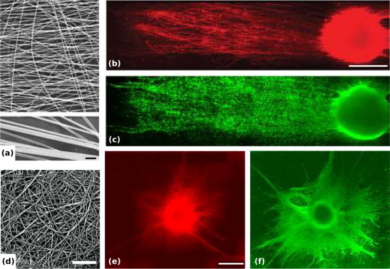
Dorsal root ganglia (DRGs) on aligned and random electrospun nanoscale fiber film in vitro. (a, d) Representative scanning electron microscopy (SEM) images of the aligned poly(acrylonitrile-comethylacrylate) fibers (a, with magnified fibers below, scale bar = 1 μm) and the random fibers (d, scale bar = 30 μm). (b, c) Double immunostained DRG on the aligned fiber film: representative montage of NF160 (a marker for axons) immunostained DRG neurons on the film (b) and montage of S-100 (a marker for Schwann cells) immunostained Schwann cells on the film (c), scale bar = 500 μm. (e, f) Double immunostained DRG on the random fiber film: representative montage of NF160+ neurons (e) and S-100+ Schwann cells (f), scale bar = 500 μm. Adapted with permission from Kim et al. (2008).