
Figure 3.
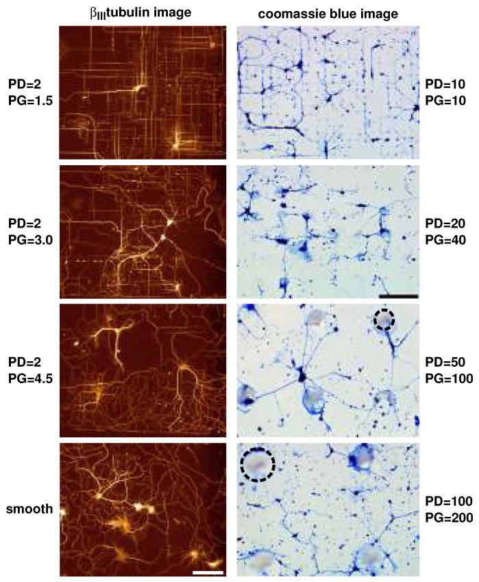
Effects of surface topography on the orientation of embryonic rat hippocampal neurons cultured on surfaces with varying pillar diameter (PD) and pillar gap (PG). Left column: Fluorescence images demonstrate the effects of poly-l-lysine-coated silicon pillars of diameter 2.0 μm at increasing pillar gap sizes. The greatest orientation was demonstrated for β III-tubulin-labeled processes on pillar gaps of 1.5 μm. Effects of orientation decreased as the gap size increased to 3.0 and 4.5 μm. Processes on smooth regions demonstrated random growth. Adapted with permission from Dowell-Mesfin et al. 2004. Right column: Optical micrographs, at the same magnification and light settings, of Coomassie blue-stained hippocampal neurons plated on glass substrates patterned with conical posts of polydimethylsiloxane (PDMS) of 10 μm diameter, 10 μm gap; 20 μm diameter, 40 μm gap; 50 μm diameter, 100 μm gap; and 100 μm diameter, 200 μm gap. Dotted circles outline representative pillars. Adapted with permission from Hanson et al. (2009).