
Figure 4.
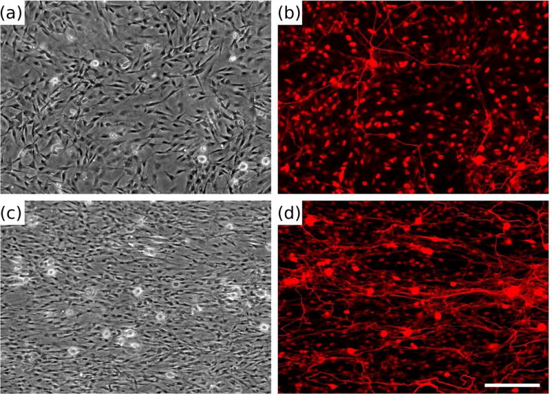
Dorsal root ganglia (DRG) morphology on biomimetic materials presenting replicated Schwann cell (SC) topography. Phase contrast (a, c) and fluorescent micrographs (b, d) of dissociated DRG cultured for 5 days on polydimethylsiloxane (PDMS) (a, b) or replicas (c, d) and stained with neurofilament immunocytochemistry (b, d). Adapted with permission from Bruder et al. (2007).