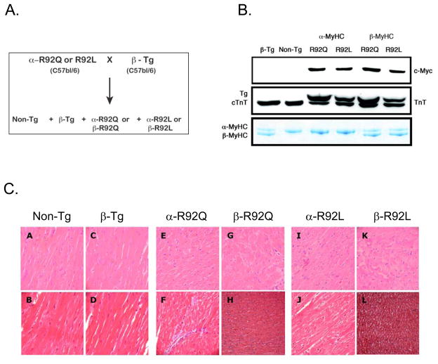
Figure 1. β-R92Q and β-R92L double transgenic mouse heart models.
A. Breeding scheme B. Protein expression levels Both R92Q and R92L mutant proteins were myc tagged at their amino termini and identified by anti-myc Western blot analysis of ventricular homogenates. Anti-Troponin T (cTnT) identified two bands, the upper band corresponding to mutant R92Q or R92L cTnT and a lower band representing endogenous cTnT. Percent expression of mutant cTnT was 67% in both α-R92Q and β-R92Q and 50% in both α-R92L andβ-R92L. Myosin heavy chain (MyHC) isoform composition was determined by glycerol containing SDS-PAGE followed by Coomassie staining. The upper band is α-MyHC while the lower band is β-MyHC. β-Tg, β-R92Q and β-R92L ventricular homogenates exhibited the same 80% β-MyHC/20% α-MyHC composition. C. Histology of Non-Tg and transgenic left ventricular sections. Hematoxylin and Eosin prepared sections are shown in upper panels and Masson’s trichrome sections are shown in lower panels. Panels A and B: Non-Tg ventricles demonstrate normal histology. Panels C and D: β-Tg ventricles also exhibited normal histology, and were free of fibrosis. Panels E and F: α-R92Q ventricles displayed myocyte disarray and mild fibrosis. Panels G and H: β-R92Q ventricles had similar histopathology to α-R92Q ventricles. Panels I and J: α-R92L ventricular sections showed myocyte disarray, but were free of fibrosis. Panels K and L: β-R92L ventricles showed similar histopathology to α-R92L ventricles. A small region of fibrosis localized to the ventricular base was observed for both β-R92Q and β-R92L hearts. Magnification = 400X for all sections.

