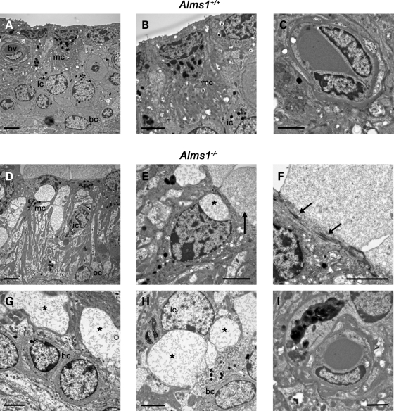
Figure 9.
Formation of membrane-bound intracellular vacuoles in Alms1−/− mice. (A–I) Transmission electron micrographs of stria vascularis from P191 Alms1+/+ and Alms1−/− mice. (A) Normal appearance of Alms1+/+ mouse stria vascularis, including marginal cells (mc), intermediate cells (ic), basal cells (bc) and a blood vessel (bv). (B) Detail of a single marginal cell. (C) Detail of a blood vessel. (D) Abnormal appearance of Alms1−/− mouse stria vascularis. Large spaces were apparent in the intermediate cell layer, and nuclei in marginal cells and basal cells were atypical in shape. (E) Detail of a single marginal cell, showing a sub-cellular vacuole (marked *) above the nucleus and a bleb extending from the apical membrane (arrow). (F) Detail of the apical membrane of a marginal cell, showing amorphous material in the apical bleb, and microfilament assemblies bordering the bleb (arrows). (G) Large membrane-bound intracellular vacuoles (*) in the intermediate cell layer, above the basal cell layer. The vacuoles contained cytoplasmic material. (H) Large membrane-bound intracellular vacuoles (*) inside an intermediate cell causing deformation of the nucleus. (I) Normal appearance of a blood vessel. Scale bars, 2 µm.

