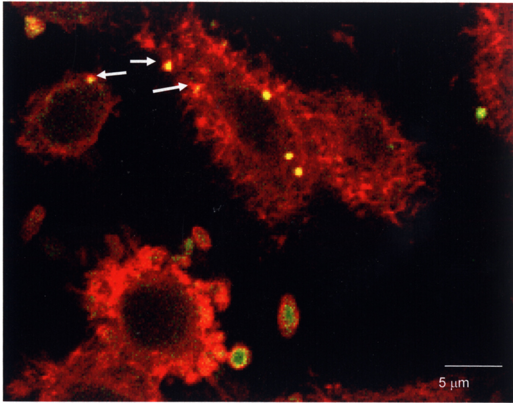
Fig. 7. Actin polymerization associated with P. gingivalis adherence and internalization by HEp-2 cells.
After infection P. gingivalis was detected with rabbit anti-RgpA adhesin domain primary antibody and Alexa Fluor 488 (green)-coupled anti-rabbit secondary antibody. Actin in HEp-2 cells was detected by staining with rhodamine phalloidin (red). Adherent external P. gingivalis cell clusters are green, internalized cells are yellow and several are surrounded by foci of polymerized actin (indicated by arrows).

