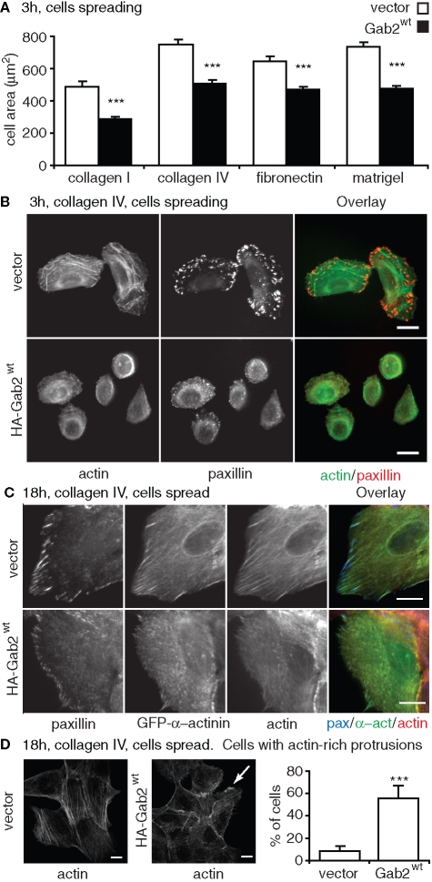
FIGURE 2:
Delayed cell spreading and impaired actin stress fiber and FA assembly in cells overexpressing Gab2. (A) Effect of Gab2 on cell spreading. The histograms indicate cell area for cells spreading for 3 h on different extracellular matrices. Values are mean ± SE of 60–80 measurements in three independent experiments. (B) Effect on focal adhesions and stress fibers during spreading. Vector controls or cells overexpressing Gab2 were plated on collagen IV for 3 h, fixed, and then stained with FITC-phalloidin or an anti-paxillin antibody. (C) Effect in spread cells. Cells were transfected with a GFP-α-actinin construct, allowed to spread for 18 h on collagen IV, and then stained for paxillin and F-actin. (D) Effect on lamellipodia and ruffles. The histograms indicate the percentage of cells with actin-rich plasma membrane protrusions (lamellipodia and ruffles), as detected by F-actin staining (representative images flank graph, arrow highlights a ruffle). Values are mean ± SD of 50 measurements in four independent experiments. *** indicates p < 0.0001 for vector vs. MCF-10A/Gab2 (by unpaired Student’s t test). All scale bars are 10 μm.

