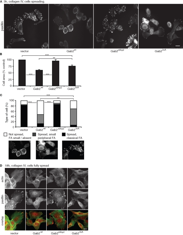
FIGURE 3:
Gab2-mediated modulation of cell spreading and cytoskeletal organization is dependent on the Shp2-binding sites. Effect on FAs (A) and cell spreading (B). The images in (A) represent paxillin staining for cells spreading for 3 h on collagen IV. In (B), the histograms indicate cell area for these cells. Values are mean ± SE of 60–80 measurements in three independent experiments. (C) Classification of cells based on FA formation and degree of spreading. More than 200 cells were classified in three independent experiments. Statistical tests compare data for spread cells exhibiting classical FAs. ** and *** indicate p < 0.001 and p < 0.0001, respectively. (D) Cytoskeletal organization and FAs in fully spread cells. Images are derived from cells allowed to spread for 18 h on collagen IV and then stained for actin or paxillin. All scale bars are 10 μm.

