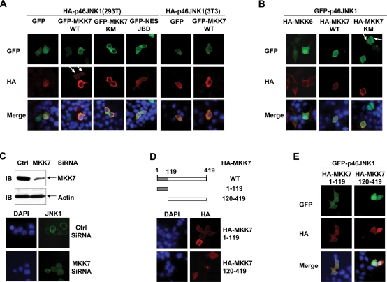
FIGURE 2:
MKK7 anchors JNK1 in the cytoplasm via its N-terminal JNK-binding domain in resting 293T and NIH3T3 cells. (A) 293T and NIH 3T3 cells were transfected with a mammalian expression vector encoding HA-p46JNK1, with cotransfection of the plasmid harboring GFP-MKK7 WT, GFP-MKK7 KM, GFP-NES-JBD, or GFP. Then 24 h after transfection, the cells were serum starved, fixed, and stained with an HA antibody. (B) 293T cells were transfected with a mammalian expression vector encoding GFP-p46JNK1, with cotransfection of the plasmid harboring HA-MKK6, HA-MKK7 WT, or HA-MKK7 KM. Then the cells were treated as described in (A). (C) 293T cells were transfected with MKK7 siRNA or the control scramble siRNA and cultured for 48 h. Cell lysates were prepared and expression of MKK7 and actin was determined by IB (Top). Ctrl, Control. The subcellular localization of endogenous JNK1 was measured as described in Figure 1A (Bottom). (D) Schematic presentation (Top) and subcellular localization (Bottom) of truncated MKK7 constructs. (E) 293T cells were transfected with a mammalian expression vector encoding GFP-p46JNK1, with cotransfection of the plasmid harboring HA-MKK7 1–119 or HA-MKK7 120–419. Then the cells were treated as described in (A). The arrows in (A) and (B) indicate that the cells without exogenous MKK7 show nuclear accumulation of exogenous JNK1, whereas the other cell in the same field with exogenous MKK7 shows cytoplasmic localization of exogenous JNK1.

