FIGURE 4:
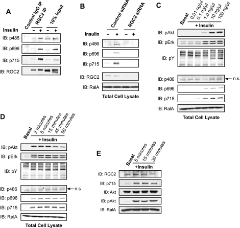
Insulin stimulates phosphorylation of RGC2 in adipocytes. (A) Serum-starved 3T3-L1 adipocytes were mock treated or stimulated with 100 nM insulin for 10 min, and lysates were immunoprecipitated using control rabbit IgG or anti-RGC2 antibody. The immune complexes from one 10-cm plate and 2% of total cell lysates were resolved on SDS–PAGE and analyzed by WB using the indicated antibodies. (B) 3T3-L1 adipocytes were transfected with control siRNA or siRNA oligos to deplete RGC2. Serum-starved cells were mock treated or stimulated with 100 ng/μl insulin for 10 min before lysis; 2% of cell lysates from a 10-cm plate of adipocytes were resolved by SDS–PAGE and subjected to WB analysis with the indicated antibodies. (C) Serum-starved 3T3-L1 adipocytes were mock treated or stimulated with insulin at the indicated doses for 10 min before lysis; 2% of cell lysates from a 10-cm plate of cells were resolved by SDS–PAGE and analyzed by WB with the indicated antibodies. (D) Serum-starved 3T3-L1 adipocytes were mock treated or stimulated with 100 ng/μl insulin for the indicated times; 2% of cell lysates from a 10-cm plate of cells were resolved by SDS–PAGE and analyzed by WB with the indicated antibodies. (E) Epididymal fat pads were excised from a mouse and starved ex vivo in serum-free DMEM for 30 min before stimulation with 100 nM insulin for the indicated times; 30 mg of protein per condition were resolved by SDS–PAGE, and RGC2 phosphorylation was determined by WB.

