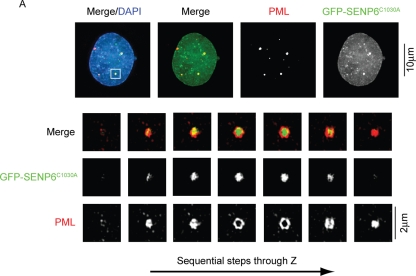
FIGURE 8:
GFP-SENP6C1030A localizes to the core domain of PML NBs. HeLa cells were transfected with GFP-SENP6C1030A 36 h before immunostaining with PML antibodies. Conventional deconvolution microscopy was performed on samples and presented as projected images. Scale bar represents 10 μm (top panels). To achieve higher resolution of the indicated PML NBs, cells were analyzed by structured illumination and are images presented as individual Z-slices. SENP6 was found to localize to the core domain in 57% of cases. Scale bars represent 2 μm.

