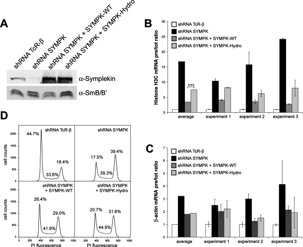
FIGURE 4:
In vivo complementation of symplekin. (A) Western blot detecting symplekin in cells depleted of TCR-β (negative control), symplekin, or in cells depleted of symplekin and simultaneously expressing Flag-tagged, RNAi-resistant wild-type or Hydro mutant symplekin. (B, C) Apparent in vivo processing efficiencies of histone H3C (B) and β-actin (C) RNA in identically treated cells. The processing efficiencies were calculated as ratios of pre-mRNAs to total RNAs and then normalized with respect to the ratio obtained in cells treated with TCR-β−specific shRNA. The less efficient histone processing complementation obtained with the symplekin Hydro mutant compared to wild-type symplekin is statistically significant (***) with a p value of 0.000027 determined by Student’s t test. (D) Cell cycle analysis by flow cytofluorometry of HeLa cells depleted of symplekin and complemented with wild-type symplekin or the symplekin Hydro mutant. The graphs represent the cell cycle distribution of cells, harvested 5 d posttransfection. The percentages of cells in the different stages of the cell cycle are indicated.

