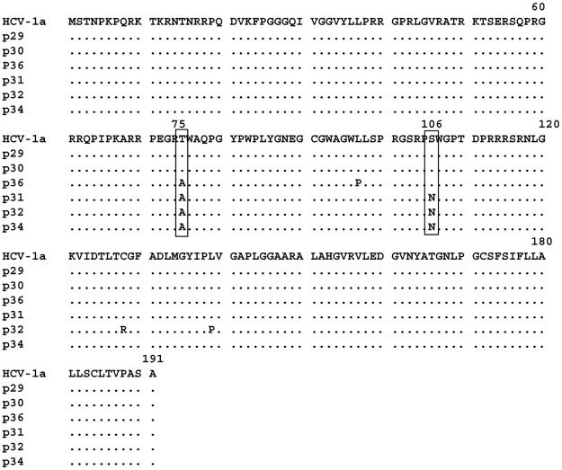
Figure 4. Amino-acid sequence alignment of the core region derived from three HCV-1a isolates positive for anti-core antibodies (p29, p30, p36) and those that were negative for anti-core antibodies (p31, p32, p34).
cDNA fragments from the HCV core region were obtained and cloned into the TOPO TA vector as described in Material and Methods. Three clones were sequenced for each patient and all three were identical for each patient. Protein sequences were analyzed using the CLUSTAL W program. The amino-acid sequence of the HCV-1a H reference strain is shown.

