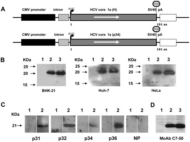
Figure 7. Expression of the HCV core protein from an anti-core negative patient (p34) in mammalian cells.
(A) Schematic representation of constructs used for the eukaryotic expression of core from the p34 HCV-1a isolate using plasmid pHPI8162 and HCV-1a H strain using plasmid pHPI 8161 (described in Material and Methods). An artificial stop codon was inserted at the 3′ end of the core gene (STOP). (B) Transient expression of the cloned HCV core gene in cultured cells. Cells were transfected with pHPI 8161 or pHPI 8162 plasmids, or the pCI empty vector. After 48 hours, cell lysates were analyzed by Western blotting using rabbit anti-core antibodies. Lane 1: cells transfected with pCI empty vector; lane 2: with a plasmid encoding HCV core (H strain); lane 3: with a plasmid encoding HCV core p34. The 21 kDA HCV core protein was detected in of all cell types. (C) The expressed core protein (M.wt. 21 KDa) is reactive with all three unusual plasma samples, p31 p32 and p34 and with a control plasma p36 positive for anti-core antibodies in all assays. NP –HCV negative plasma; extract from cells transfected with pCI control plasmid (Lane 1) and from cells transfected with a plasmid encoding HCV core p34 (Lane 2). (D) The expressed core protein is reactive with anti-core MAbC7-50 that recognizes an epitope localized in the aa(21–57) sequence. Extract from cells transfected with the pCI control plasmid (Lane 1), with a plasmid encoding HCV core from H strain (Lane 2) and with a plasmid encoding HCV core from p34 (Lane 3).

