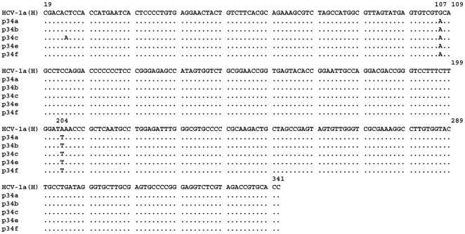
Figure 10. Nucleotide sequence alignment of the cDNA sequence that corresponds to the 5′UTR region from patient p34, negative for anti-core and positive for anti-core+1/ARFP antibodies.
The c-DNA fragments were obtained by PCR and cloned into the HincII site of pUC19 vector. Five clones were isolated and sequenced. The nucleotide sequence of HCV1a H strain is shown.

