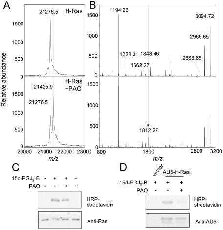
Figure 4. PAO binds to the C-terminal peptide of H-Ras in vitro and in cells.
(A) Full length H-Ras was incubated with PAO and analyzed by MALDI-TOF MS. (B) Control or PAO-treated H-Ras was subjected to tryptic digestion and the resulting peptides analyzed by MALDI-TOF MS. The peak of m/z 1812.27, corresponding to the formation of an adduct between PAO and the K170-K185 peptide, is marked by an asterisk. Dotted lines mark the positions equivalent to those of the modified species in the untreated samples. (C) H-Ras at 5 µM was incubated with 50 µM PAO for 30 min before addition of 1 µM biotinylated 15d-PGJ2 (15d-PGJ2-B) for 1 h. (D) COS-7 cells were transiently transfected with empty vector or a plasmid coding for AU5-H-Ras. 24 h after transfection cells were pre-treated with vehicle or 1 µM PAO for 90 min before incubation with 5 µM 15d-PGJ2-B for 90 min. Incubation mixtures from (C) and cell lysates from (D) were analysed by SDS-PAGE followed by transfer to membrane and detection of incorporated biotin with horseradish peroxidase (HRP)-streptavidin and of the Ras protein by immunoblot with anti-pan Ras or anti-AU5 antibody and enhanced chemiluminescence (ECL). Results shown are representative of at least three assays.

