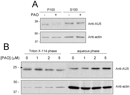
Figure 5. Effect of PAO on H-Ras subcellular localization.
(A) COS-7 cells were transiently transfected with expression vectors coding for AU5-H-Ras wt. After serum starvation for 16 h, cells were treated with vehicle or 1 µM PAO for 1 h. Cells were lysed and postnuclear supernatants were separated into S100 and P100 fractions by ultracentrifugation at 200,000× g for 30 min. (B) AU5-H-Ras transfected cells were treated with the indicated concentrations of PAO for 90 min and cell lysates were subjected to fractionation in Triton-X114. The amount of AU5-H-Ras present in the various fractions was estimated by western blot with anti-AU5 antibody (upper panels). The upper component of the H-Ras doublet is indicated by an arrowhead. Levels of actin were assessed as control (lower panels). Blots shown are representative of three assays.

