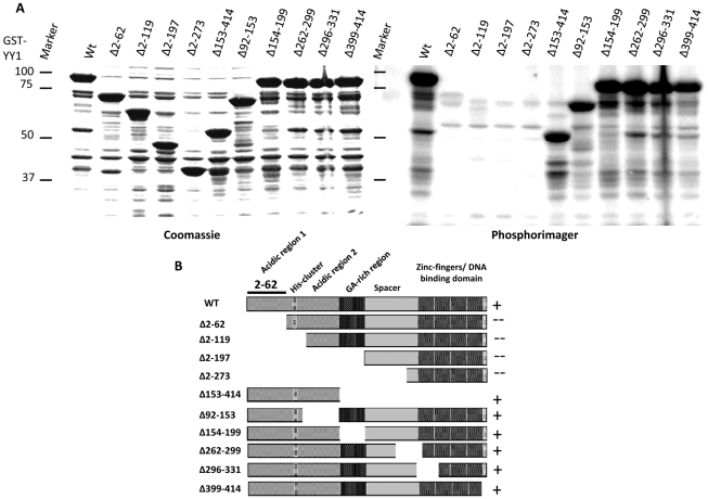
Figure 2. Plk1 phosphorylates YY1 in the N-terminal activation domain in vitro.
(A) Coomassie blue staining (left) and phosphorimager exposure (right) of SDS-PAGE gel analysis of the radioactive in vitro kinase assay reactions using purified Plk1 and a panel of GST-tagged YY1 deletion mutants. The specific YY1 deletions are indicated above the lanes. Equal amounts of purified Plk1 were added to all reactions. (B) Diagram of the deletion mutants of YY1 used in the kinase assay in (A). Evidence of phosphorylation shown in (A) is indicated by the (+) sign, whereas the absence of evidence of phosphorylation is indicated by a (--) sign. The region identified as the site for phosphorylation by Plk1 is indicated (amino acid residues 2–62).

