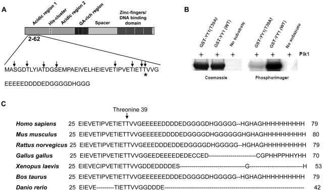
Figure 3. Plk1 phosphorylates YY1 at threonine 39 in vitro.
(A) Diagram displaying the different domains of the YY1 protein. Amino acid residues 2–62 are shown; serine and threonine residues in amino acids 2–62 are indicated by arrows. The predicted phosphorylation site at threonine 39 is indicated with a star. (B) Coomassie blue staining and phosphorimager exposure of the SDS-PAGE gel analysis of the radioactive in vitro kinase assay reactions. Kinase reactions include Plk1 only (no substrate lane), Plk1 with GST-YY1 wild type (WT) or mutant (T39A). (C) Amino acid sequence alignment of the N-terminal domain of the YY1 protein from different species, as indicated.

