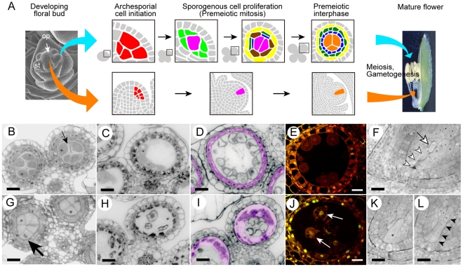
Figure 1. Developmental aberration of germline and nursery cells in the mel2 mutant.
(A) A schematic illustration of the germline cell development in the anther and ovule in rice. Red-colored cells indicate the archesporial cells; magenta, sporogenous cells; light green, primary parietal cells; yellow, endothecium; brown, secondary parietal cells; dark green, middle layer cells; blue, tapetal cells; mandarin, meiocytes at premeiotic interphase. (B–L) Plastic sections of wild-type (B–F) and mel2 (G–L) reproductive organs during sporogenesis. (B, G) Premeiotic interphase. The callose accumulation among PMCs (a small arrow) was absent in the mutant. A large arrow indicates asynchronous, aberrant mitosis of the PMC. (C, H) Meiotic I prophase (diakinesis). (D, I) Post-meiotic microspore stage. Tapetal cells were pseudo-colored in magenta. (E, J) TUNEL assay. White arrows indicate TUNEL signals representing apoptotic DNA fragmentation found in nuclei of disrupted PMCs. (F, K, L) Ovules at post-meiosis. Three of tetrad spores (open arrowheads) were degenerated, and a single megaspore (an open arrow) underwent megagametogenesis after the completion of wild-type meiosis, whereas meiosis progression was stagnant at various steps in the mutant (K, L). Closed arrowheads indicate an equal size of tetrad spores. Bars, 10 µm.

