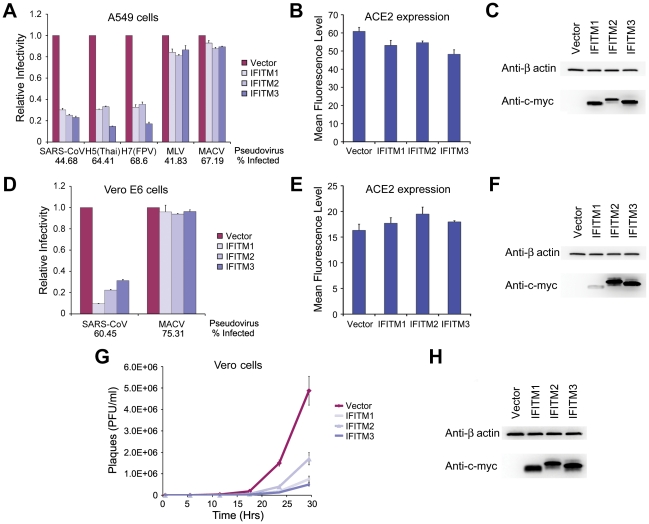
Figure 3. SARS-CoV S infection is restricted by IFITM1, 2, and 3.
(A) A549 or (D) Vero E6 cells transduced to express ACE2 were subsequently transduced to express the indicated c-myc-tagged IFITM proteins or with vector alone. Two days later, cells were infected with indicated pseudoviruses. Pseudovirus infection was determined by flow cytometry, and normalized to that of cells transduced with vector alone. Differences in pseudovirus entry between vector alone and IFITM expressing cells are significant (P<0.05) for all SARS-CoV and IAV pseudoviruses. In parallel, cell-surface expression of ACE2 was assayed using aliquots of the same (B) A549 or (E) Vero E6 cells analyzed in (A) and (D), respectively. Cells were labeled with Alexa-488 conjugated S protein RBD of SARS-CoV and analyzed by flow cytometry. ACE2 expression is shown as mean fluorescence intensity. Expression of c-myc-tagged IFITM proteins was assayed by western blot using aliquots of the same (C) A549 or (F) Vero E6 cells analyzed in (A) and (D), respectively. Each panel represents at least three experiments with similar results. (G) Vero cells transduced to express indicated c-myc-tagged IFITM proteins or with vector alone were incubated in duplicate with infectious SARS-CoV at a MOI of 0.1 for 1 hour. Supernatants were harvested 1, 6, 12, 18, 24, or 30 hours later and viral titers were measured by plaque assay. (H) Expression of c-myc-tagged IFITM proteins was assayed by western blot using aliquots of cells analyzed in (G).

