Fig. 5.
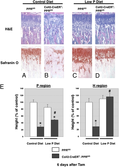
Concurrent administration of a low-phosphate diet prevents disappearance of the growth plate. H&E (Upper) and Safranin O (Lower) staining of longitudinal cross-sections of tibiae in PPRfl/fl (A and C) and Coll2-CreERT:PPRfl/fl (B and D) mice at 4 wk of age at 6 d after Tam administration. PPRfl/fl (A and C) and Coll2-CreERT: PPRfl/fl (B and D) mice were fed a control diet (A and B) or a low-phosphate (P) diet (C and D) from 28 to 35 d of age. (E) Statistical analysis of the length of the columnar proliferating (Left) and hypertrophic (Right) regions of the proximal growth plate of the tibiae in PPRfl/fl and Coll2-CreERT:PPRfl/fl mice fed a control or P diet. White bars represent PPRfl/fl mice, and black bars represent Coll2-CreERT:PPRfl/fl mice. *P < 0.05, significantly different from PPRfl/fl mice fed a control diet; #P < 0.05, significantly different from the value obtained in Coll2-CreERT:PPRfl/fl mice fed a control diet. Data are representative of experiments performed on sections from three or more mice for each condition.

