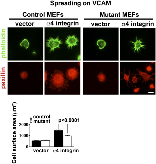
Fig. 4.
Abi1 mutant MEFs expressing α4 integrin show diminished cell spreading on VCAM1. Control (Abi1+/−/Abi2+/−) or mutant (Abi1−/−/Abi2+/−) MEFs were transduced with vector alone or vector containing WT full-length α4 integrin. Cells were sorted by FACS to obtain cell populations expressing equivalent amounts of α4, and these cells were plated immediately on coverslips coated with VCAM1. After 2 h, cells were fixed and processed for indirect immunofluorescence for detection of paxillin (red). Actin staining with phalloidin is shown (green). Quantification of cell spreading was analyzed by measuring cell surface area of phalloidin stained cells using Zeiss software (Bottom). Abi1 mutant cells expressing α4 integrin exhibit significantly reduced spreading compared with their control counterparts on VCAM1 substratum. (Scale bar: 10 μm.)

