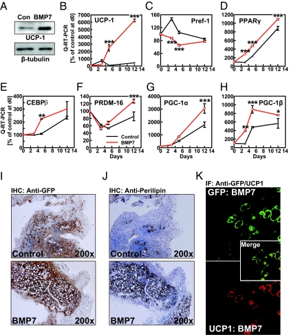
Fig. 2.
Pretreatment of muscle-resident ScaPCs with BMP7 results in formation of brown adipocytes in vitro and in vivo. (A) Protein levels of UCP1 following pretreatment with BMP-7 and 48 h of adipogenic induction. (B–H) Quantitative RT-PCR analysis of gene expression during differentiation of ScaPCs: 16 h after seeding cells (day 0), ScaPCs were pretreated with 3.3 nM BMP7 for 3 d (day 3), followed by adipogenic induction for 48 h (day 5) and 7 d of adipogenic maturation (day 12). Expression of (B) UCP1, (C) preadipocyte factor-1 (pref-1), (D) PPARγ, (E) C/EBPβ, (F) PRDM16, (G) PGC-1α, and (H) PGC-1β. Red lines depict expression levels for BMP-7 treated cells, and controls cells are indicated in black. Data are presented as mean ± SEM (n = 3). (I) GFP immunohistochemical staining in skeletal muscle of non-GFP isogenic recipient mice transplanted with ScaPCs treated with BMP7 or vehicle for 4 d (original magnification: 200×). (J) Perilipin immunohistochemical staining of a tissue section adjacent to the section stained for GFP, as shown in panel I (original magnifications 200×). (K) Immunofluorescence detection of colocalization of GFP expression with UCP1 expression. (Upper) Detection of GFP (green); (Lower) expression of UCP1 (red). Merge (Inset) demonstrates colocalization of GFP and UCP1 (yellow). Sections adjacent to those showing GFP-positive staining in J were used for IF (original magnification: 400×). *P < 0.05; **P < 0.01; ***P < 0.001.

