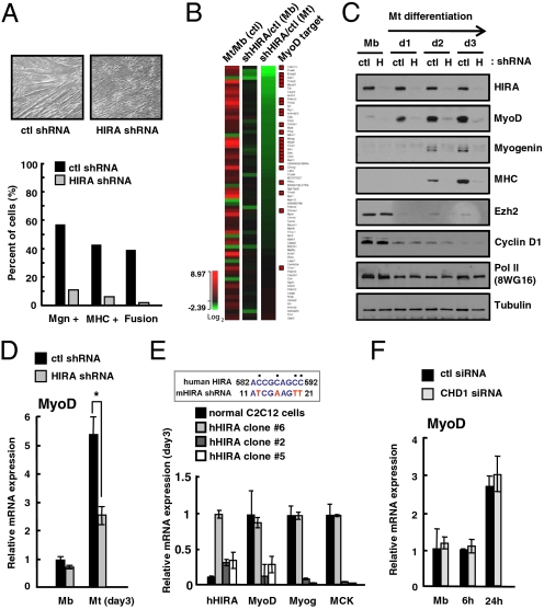
Fig. 2.
HIRA is an upstream regulator of MyoD and essential for skeletal myogenesis. (A) Myotube formation of control shRNA (ctl) or shHIRA C2C12 cells evaluated by phase contrast microscopy. Stable polyclonal shHIRA cells that express reduced HIRA were analyzed for myogenic differentiation. Myogenin+, MHC+ cells, and the extent of cell fusion were analyzed by immunofluorescence microscopy. (B) Genes known to be involved in muscle differentiation were selected and their expression changes were compared in the ctl and shHIRA cells. The heat map shows genes sorted in the order of the effect of HIRA depletion in myotubes; shHIRA/ctl (Mt). Genes reported as MyoD targets are marked by red squares on the right column. (C) The protein level of individual genes was monitored by immunoblotting analysis of whole cell lysate from ctl or shHIRA (H) cells on each differentiation day. Ezh2, Cyclin D1; myogenesis markers, pol II, Tubulin; loading controls. (D) Relative mRNA level of MyoD in control (ctl) and shHIRA cells analyzed by quantitative RT-PCR. Error bars represent SD, n = 3. The significance of difference was evaluated (* P value < 0.05). (E) shHIRA C2C12 cells were rescued by human HIRA. Three independent clones that express different levels of supplemented human HIRA were selected for analysis. Control C2C12 and three clones were subjected to quantitative RT-PCR for the indicated genes. The expression of human HIRA was monitored by human-specific primers. The approximate expression level of human HIRA of clone #6 was estimated by immunoblotting assay as ∼60% of that of mouse HIRA of control cells (Fig. S5A). (Upper) Part of the nucleotide sequence of human HIRA that deviates from the shRNA used for targeting mouse HIRA. (F) Quantitative RT-PCR analysis of MyoD mRNA levels in control (ctl) or siCHD1-treated C2C12 cells at each differentiation time point.

