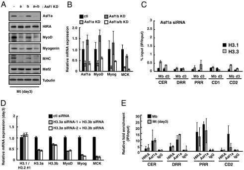
Fig. 4.
H3.3 incorporation is mediated by HIRA/Asf1a for MyoD activation. (A and B) The expression level of each gene was analyzed by immunoblotting (A) or quantitative RT-PCR analysis (B) after cells were shifted to the differentiation condition for 3 d (day3). To knock down Asf1a and Asf1b separately, either nontargeting control siRNA or Asf1a-specific siRNA was transfected into C2C12 cells expressing control (lanes 1,2) or Asf1b-specific shRNA (lanes 3,4) (Fig. S7A). (C) ChIP was performed to monitor incorporation of eH3 at MyoD regulatory and coding regions in Asf1a siRNA-treated C2C12 cells. ChIP was analyzed as described in Fig. 3. (D) The assessment of the relative mRNA levels of myogenic markers in C2C12 cells treated with a mixture of siRNAs targeting H3.3a and H3.3b transcripts. Control and siH3.3-treated cells were induced to differentiate for 3 d and subjected to quantitative RT-PCR for the analysis of the indicated genes. (E) Relative occupancy of HIRA and Asf1a was analyzed by ChIP. Chromatin prepared from myoblasts or myotubes was immunoprecipitated with anti-HIRA(WC15), anti-Asf1a or normal IgG as a control. Acquired signals were normalized to input and IgG. Error bars represent SD, n = 2 or more.

