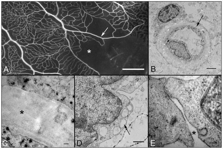
Figure 4.
Higher magnification view of the region of retina shown in Figure 1 examined using electron microscopy showing capillary dropout (asterisk) and arterial pruning (arrow). Electron micrograph of a viable capillary (B and C) from the region shown in “A”. The electron dense particles in the endothelial cytoplasm are lead reaction product from the ADPase reaction. The endothelial ultrastructure appears intact with characteristic nucleus, plasmalemma vesicles and cytoplasmic inclusions. A pericyte is seen in the close contact with the basal lamina (B) but the granular appearance of this cell and poorly defined cytoplasmic membrane may represent postmortem changes. The basal lamina is thickened and amorphous (asterisk in “C”). Endothelial cells sometimes were vacuolated (arrow in D) and open cell junctions were occasionally observed (asterisk in E). The majority of the intercellular junctions lacked the distinct zonula occludens (tight junction), which tend to be extensive in healthy retinal vessels. Scale bar = 1mm in A, 2 microns in B, 100nm in C, 500nm in D, 100nm in E.

