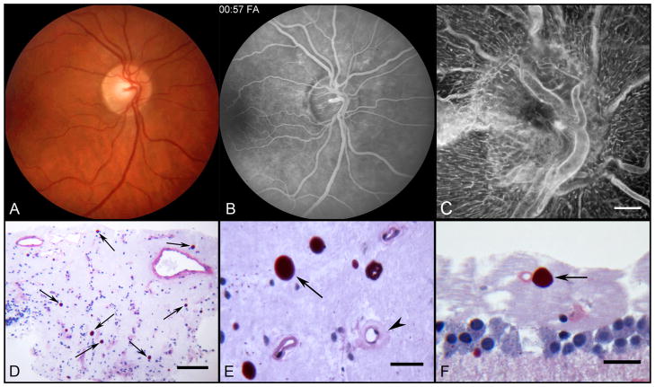
Figure 5.
Color fundus photograph (A) of the right fundus shows patchy irregular caliber of the inferonasal branch retinal artery and of the small macular arteries and veins. Venous phase fluorescein angiogram (B) of the same field shows better vascular detail and telangiectasis of the optic nerve head. Note patchy abnormal retinal hyperfluorescence superior to the optic nerve corresponding to leaking capillaries. ADPase flat embedded optic nerve head (C) and peripapillary retina of the right eye (scale bar = 300 μm). Section through nerve head (D) showing numerous corpora amylacea (arrows) stained with PAS (scale bar = 100 μm). Higher magnification of corpora amylacea (E) in nerve head (arrow) near a capillary (arrowhead) with an abnormally thickened wall (scale bar = 20 μm). Corpora amylacea (arrow) in the nerve fiber layer (F) of the paramacular retina. Scale bar = 20 μm (D–F. PAS and hematoxylin).

