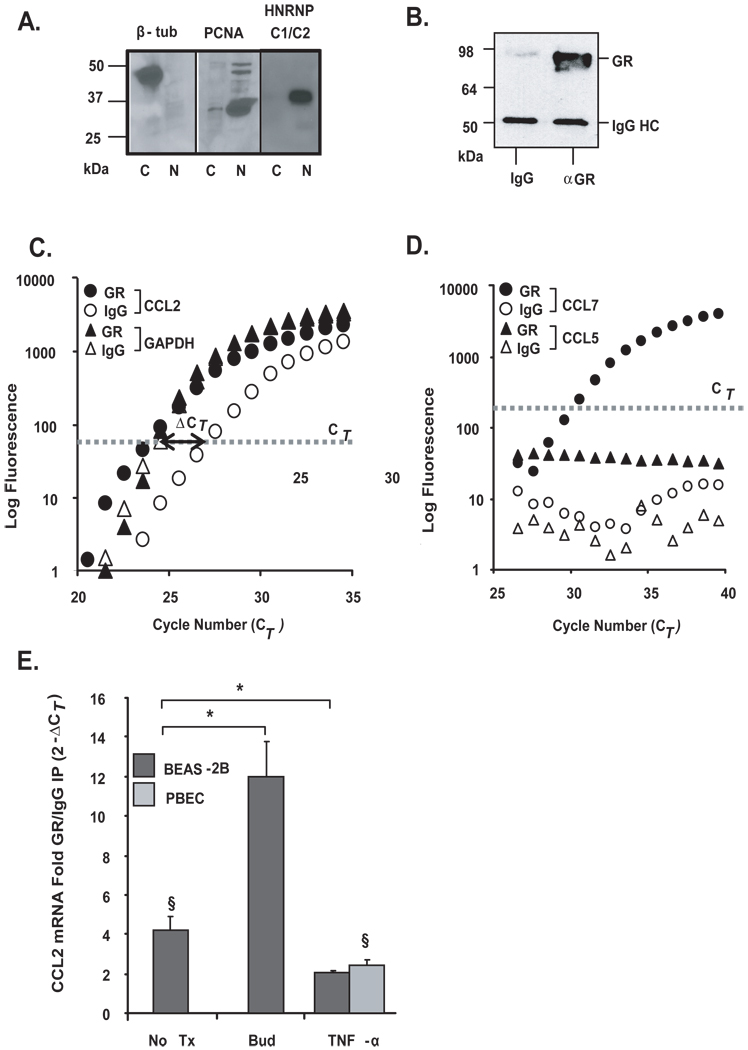
Figure 3. Association of chemokine transcripts with GR in airway epithelial cells.
A. The purity of cytoplasmic lysates was verified by Western blot analysis of a cytoplasmic protein [β-tubulin (β-tub)] and nuclear proteins (PCNA, HNRNP C1/C2), detected exclusively in cytoplasmic (c) and nuclear (n) extracts, respectively. B. Western Blot analysis of GR after immunoprecipitation (IP) of protein-mRNA complexes obtained using the monoclonal anti-GR antibody (αGR) or the IgG-control antibody control (IgG), showing selectivity of the IP. Bands corresponding to GR and IgG heavy chain (IgG HC) are indicated. C. Real-time PCR amplification plot of CCL2 and GAPDH mRNA (representative of n=10) and D. of CCL7 and CCL5 mRNA (representative of n=5), both after GR (filled circles) or IgG control (open circles) IP. The bold line with arrow points indicates the difference in Ct (ΔCT) between the CCL2 mRNA detected in the GR-IP versus the IgG-IP. E. Mean ± SEM fold GR/IgG IP enrichment for CCL2 mRNA (expressed as 2−ΔCT) in BEAS-2B and PBEC cells unstimulated (n=10) or treated with budesonide (bud, n=4) or TNFα (n=3). * p < 0.03 for treated samples vs. untreated, § p < 0.03 vs. GR-IP control.

