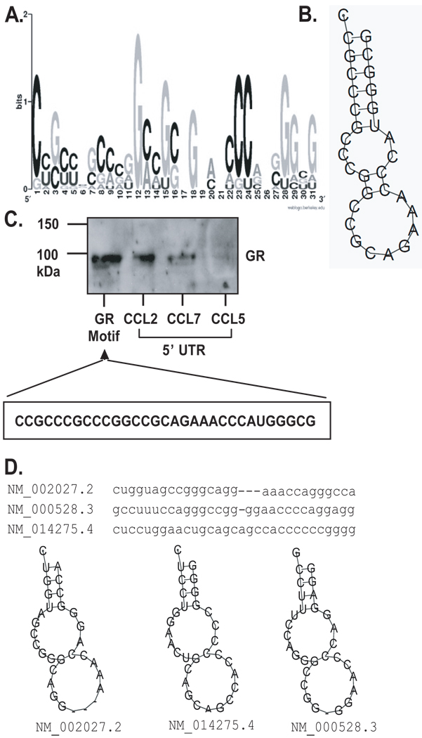
Figure 7. Primary sequence and secondary structure of the predicted GR mRNA motif.
A. Graphic logo representing the probability matrix of the GR motif, showing the relative frequency of each nucleotide for each position within the motif sequence. The motif is originated from the experimental data set from the array study. B. Secondary structure of the GR motif comprising the nucleotides with highest frequency for each position within the motif shown in A. C. Biotin pull-down assay showing association of GR from unstimulated BEAS-2B cell lysates with the GR motif shown in B., compared to GR association with the full-length 5’UTR of CCL2, CCL7 and CCL5 mRNA (the latter as negative control). D. Sequence and secondary structure of the GR motif in three mRNAs from the UniGene list of putative GR targets; the corresponding RefSeq accession numbers and names are shown.

