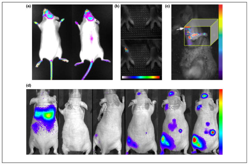
Figure 2.

Applications of musculoskeletal molecular imaging. (a) Two representative images of female OC-FLuc transgenic mice at 6–8 weeks of age. Bioluminescence imaging could detect osteocalcin activity in areas of active bone remodeling, including calvaria bones, teeth area, paws and proximal tail vertebra e. Images courtesy of Dr. Yoram Zilberman, Skeletal Biotech Laboratory (Professor Dan Gazit), Hebrew University-Hadassah Medical Center. (b) Imaging macrophage infiltration into inflamed joints using a NIR fluorophore-conjugated folate. The top panel shows a white-light image and the bottom panel the merged fluorescence image in which the onset of inflammation was detected 30 h after induction of arthritis in the right wrist. The color bar indicates signal intensity ranging from low (white) to high (red). Reproduced with permission from [18]. (c) Fluorescence molecular tomography of bone formation at the mineralization stage utilizing a NIR fluorophore-tagged bisphosphonate. The image shown was acquired 3 weeks after transfer of BMP-2-enhanced MSCs into a radial bone defect and showed a signal along the defect site. The shoulder of the animal is marked by an arrow. The color bar indicates signal intensity ranging from low (violet) to high (red). Reproduced with permission from [72]. (d) Temporal-spatial homing of macrophages to a bone cement particle-challenged femur was revealed by a series of bioluminescence images taken every 48 h over 10 days (from left to right). The color bar indicates signal intensity ranging from low (violet) to high (red). Reproduced with permission from [73].
