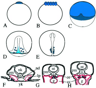
Figure 1.
Schematic representation of the migration of germ cells in medaka embryos (see also ref. 24). Olvas expressing cells or regions are colored blue (dark blue indicates more intense expression). Lateral plate mesoderm and coelomic epithelium are shown in red. (A) 1 cell (stage 2); (B) morula (stage 8); (C) early gastrulation (stage 13); (D) late gastrulation (stage 16); (E) 4 somites (stage 20); (F) stage 25; (G) stage 35 (F–H, cross sections). cc, ceolomic cavity; ce, coelomic epithelium; g, gut; lp, lateral plate mesoderm; nd, nepheric duct; n, notocord; nk, neural keel; nt, neural tube; pm, paraxial mesoderm; yk, yolk.

