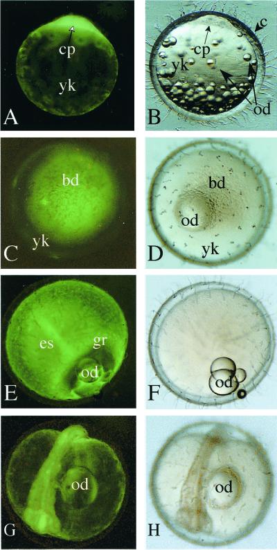
Figure 6.
Maternal expression of GFP fluorescence during embryogenesis. (A, C, E, and G) Fluorescent images. (B, D, F, and H) Bright field images. (A and B) One-cell stage, lateral view. The cytoplasm of a single cell shows GFP fluorescence. (C and D) Late morula (stages 11 and 12, animal pole view). (E and F) Gastrulation (stage 16, lateral view). (G and H) Otic vesicle formation stage (stage 21, five somites). bd, blastoderm; c, chorion; cp, cytoplasm; es, embryonic shield; gr, germ ring; od, oil droplet; yk, yolk.

